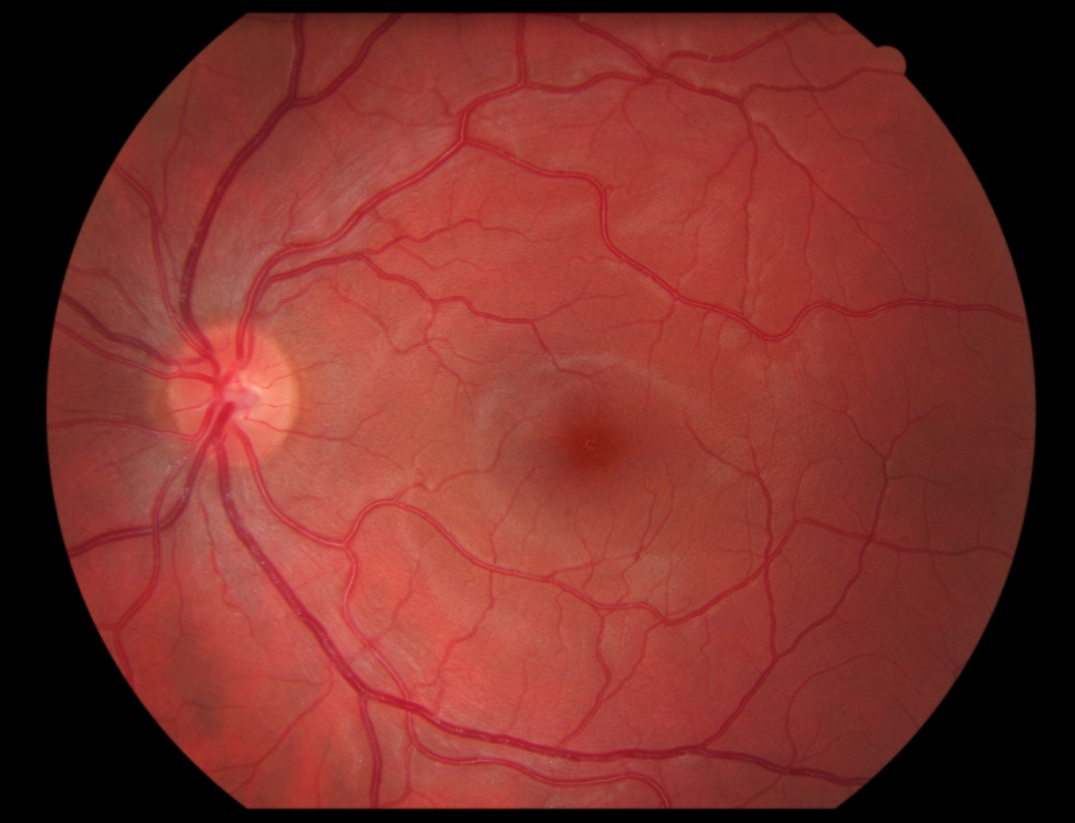
Figure 1. [The normal human retina fundus]. - Webvision - NCBI
Por um escritor misterioso
Last updated 15 julho 2024
![Figure 1. [The normal human retina fundus]. - Webvision - NCBI](https://www.ncbi.nlm.nih.gov/books/NBK554706/bin/Archetecture_Fovea-Image006.jpg)
The normal human retina fundus photo shows the optic nerve (right), blood vessels and the position of the fovea (center).
![Figure 1. [The normal human retina fundus]. - Webvision - NCBI](https://www.pnas.org/cms/10.1073/pnas.2307380120/asset/a3533755-1d49-4826-ba92-7697defec4a7/assets/images/large/pnas.2307380120fig08.jpg)
Cellular migration into a subretinal honeycomb-shaped prosthesis for high-resolution prosthetic vision
![Figure 1. [The normal human retina fundus]. - Webvision - NCBI](https://pub.mdpi-res.com/applsci/applsci-08-00155/article_deploy/html/images/applsci-08-00155-g003.png?1569808293)
Applied Sciences, Free Full-Text
![Figure 1. [The normal human retina fundus]. - Webvision - NCBI](http://webvision.org.es/wp-content/uploads/2018/01/Myopia-Fig1.jpg)
The Science Behind Myopia. Brittany J. Carr and William K. Stell - Webvision
![Figure 1. [The normal human retina fundus]. - Webvision - NCBI](https://www.ncbi.nlm.nih.gov/books/NBK11556/bin/factsf5.gif)
Facts and Figures Concerning the Human Retina - Webvision - NCBI Bookshelf
![Figure 1. [The normal human retina fundus]. - Webvision - NCBI](https://www.ncbi.nlm.nih.gov/books/NBK482309/bin/retinal_degeneration-Image044.jpg)
Figure 38. [Summary figure of the normal]. - Webvision - NCBI Bookshelf
![Figure 1. [The normal human retina fundus]. - Webvision - NCBI](https://media.springernature.com/lw685/springer-static/image/chp%3A10.1007%2F978-3-030-25886-3_22/MediaObjects/436773_1_En_22_Fig1_HTML.png)
Image Analysis for Ophthalmology: Segmentation and Quantification of Retinal Vascular Systems
![Figure 1. [The normal human retina fundus]. - Webvision - NCBI](https://www.ncbi.nlm.nih.gov/books/NBK1222/bin/retinoschisis-Image001.jpg)
Figure 1. [Fundus photo of a male]. - GeneReviews® - NCBI Bookshelf
![Figure 1. [The normal human retina fundus]. - Webvision - NCBI](http://webvision.med.utah.edu/wp-content/uploads/2011/01/OCTmacula.jpg)
Simple Anatomy of the Retina by Helga Kolb – Webvision
![Figure 1. [The normal human retina fundus]. - Webvision - NCBI](https://media.springernature.com/m685/springer-static/image/art%3A10.1007%2Fs11042-020-09041-y/MediaObjects/11042_2020_9041_Fig1_HTML.png)
Computerized retinal image analysis - a survey
![Figure 1. [The normal human retina fundus]. - Webvision - NCBI](https://media.springernature.com/m685/springer-static/image/art%3A10.1186%2Fs13024-023-00655-y/MediaObjects/13024_2023_655_Fig4_HTML.png)
Retinal ganglion cell repopulation for vision restoration in optic neuropathy: a roadmap from the RReSTORe Consortium, Molecular Neurodegeneration
![Figure 1. [The normal human retina fundus]. - Webvision - NCBI](https://eophtha.com/images/uploads/15974739483541657755f37849ce2a63.jpg)
Anatomy of Retina
Recomendado para você
-
Retina - American Academy of Ophthalmology15 julho 2024
-
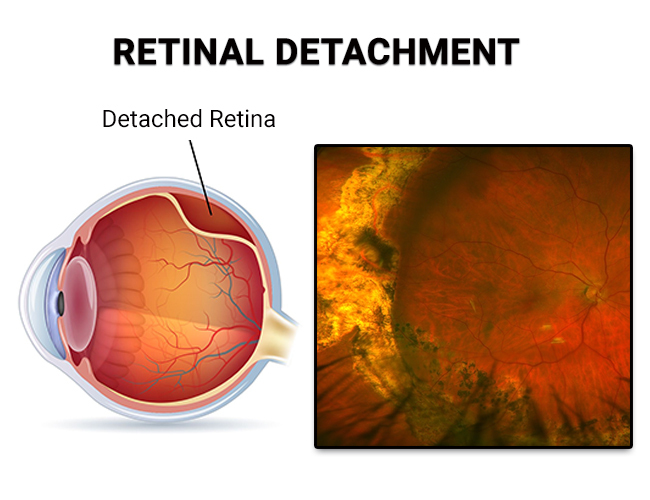
 What is the Retina? Retinal detachment and other retinal issues.15 julho 2024
 -
 Descolamento de Retina - Instituto de Moléstias Oculares15 julho 2024
 -
 Retina Specialist - Why should I see one? - SK Retina15 julho 2024
 -
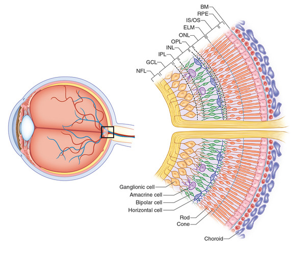
 The retina and retinal pigment epithelium (RPE)15 julho 2024
 -
 Eyeball In Section Structure Of The Retina Closeup Stock15 julho 2024
 -
 Retina Problems: Warning Signs You May Have a Retinal Disease15 julho 2024
 -
 Retinal Detachment NYC - Vitreous Retina Macula Consultants of New15 julho 2024
 -
 Retina Imaging Village Optical15 julho 2024
 -
 Normal Retinal Anatomy - The Retina Reference15 julho 2024
você pode gostar
-
 Um Jogo É Jogado No Tabuleiro De Xadrez. Parte Das Figuras Fica Ao Lado Do Tabuleiro. Cavalo Preto No Campo Do Centro. Posição Complexa, Combinação. Considere O Próximo Passo. Fotos, retratos, imágenes15 julho 2024
 -
 Brinquedo Infantil Galinha Pintadinha Pianinho Bate e Toque Yes Toys 2022415 julho 2024
 -
 O retorno do 'Messi Careca': Guarani anuncia Régis como reforço15 julho 2024
 -
 Reverse Shield Minecraft Texture Pack15 julho 2024
 -
Conjunto SHEIN lilás 🦋15 julho 2024
-
 Deatte 5-byou de Battle - Dublado – Episódio 10 Online - Hinata Soul15 julho 2024
 -
me when i halt15 julho 2024
-
 Chinese animationMo Dao Zu Shi comes to Japan! Based on the BL15 julho 2024
 -
 Chess world Cartoon Boris Spassky15 julho 2024
 -
 SLASH THE COLLECTION15 julho 2024