Morphology of Leydig cells in the testes after in vivo MCP-1 treatment.
Por um escritor misterioso
Last updated 15 julho 2024

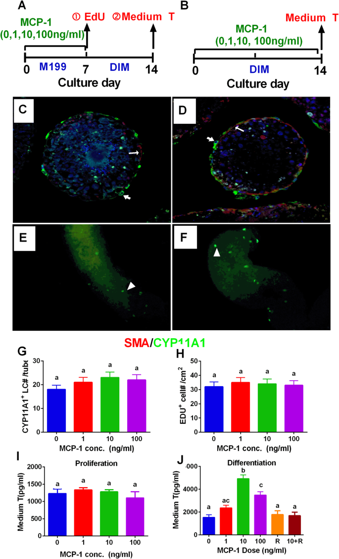
Monocyte Chemoattractant Protein-1 stimulates the differentiation of rat stem and progenitor Leydig cells during regeneration, BMC Developmental Biology

Testicular macrophages are recruited during a narrow time window by fetal Sertoli cells to promote organ-specific developmental functions
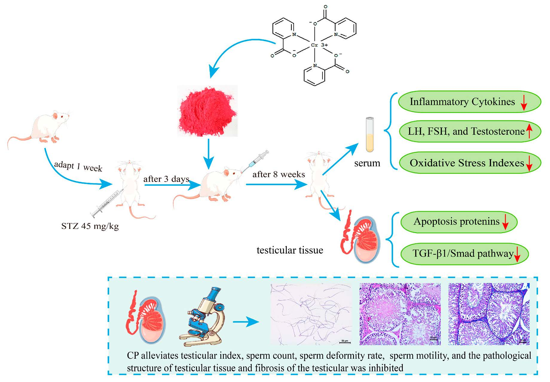
Molecules, Free Full-Text
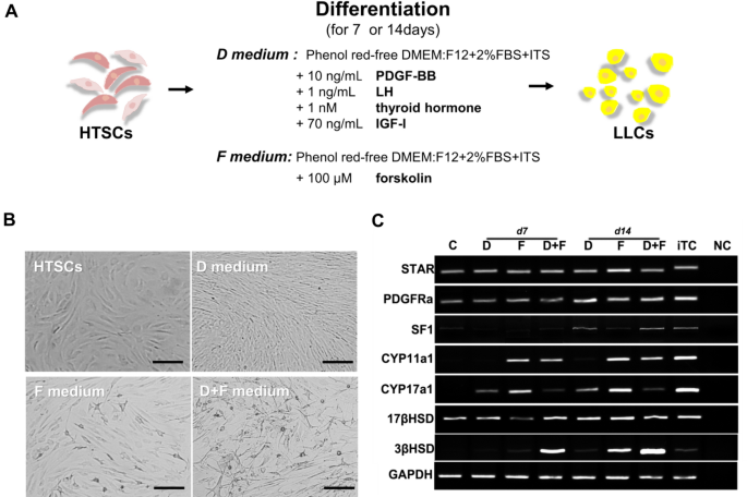
Rapid Differentiation of Human Embryonic Stem Cells into Testosterone-Producing Leydig Cell-Like Cells In vitro
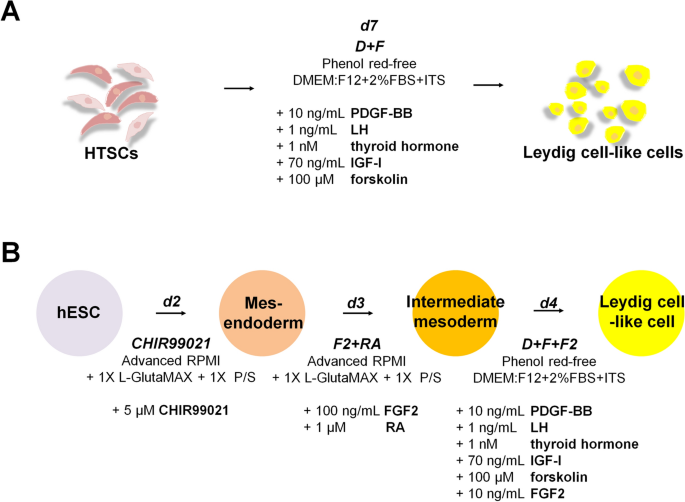
Rapid Differentiation of Human Embryonic Stem Cells into Testosterone-Producing Leydig Cell-Like Cells In vitro

Stem Leydig cells: Current research and future prospects of regenerative medicine of male reproductive health - ScienceDirect
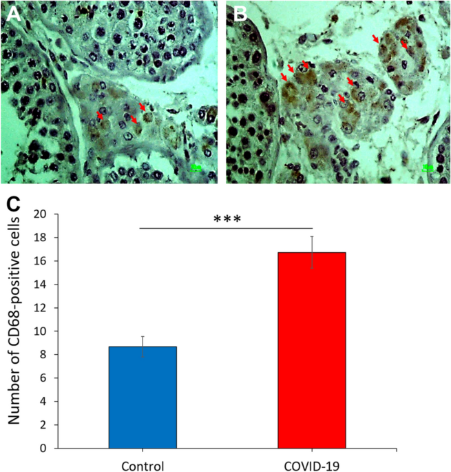
COVID-19 disrupts the blood–testis barrier through the induction of inflammatory cytokines and disruption of junctional proteins

Stem Leydig cells: Current research and future prospects of regenerative medicine of male reproductive health - ScienceDirect
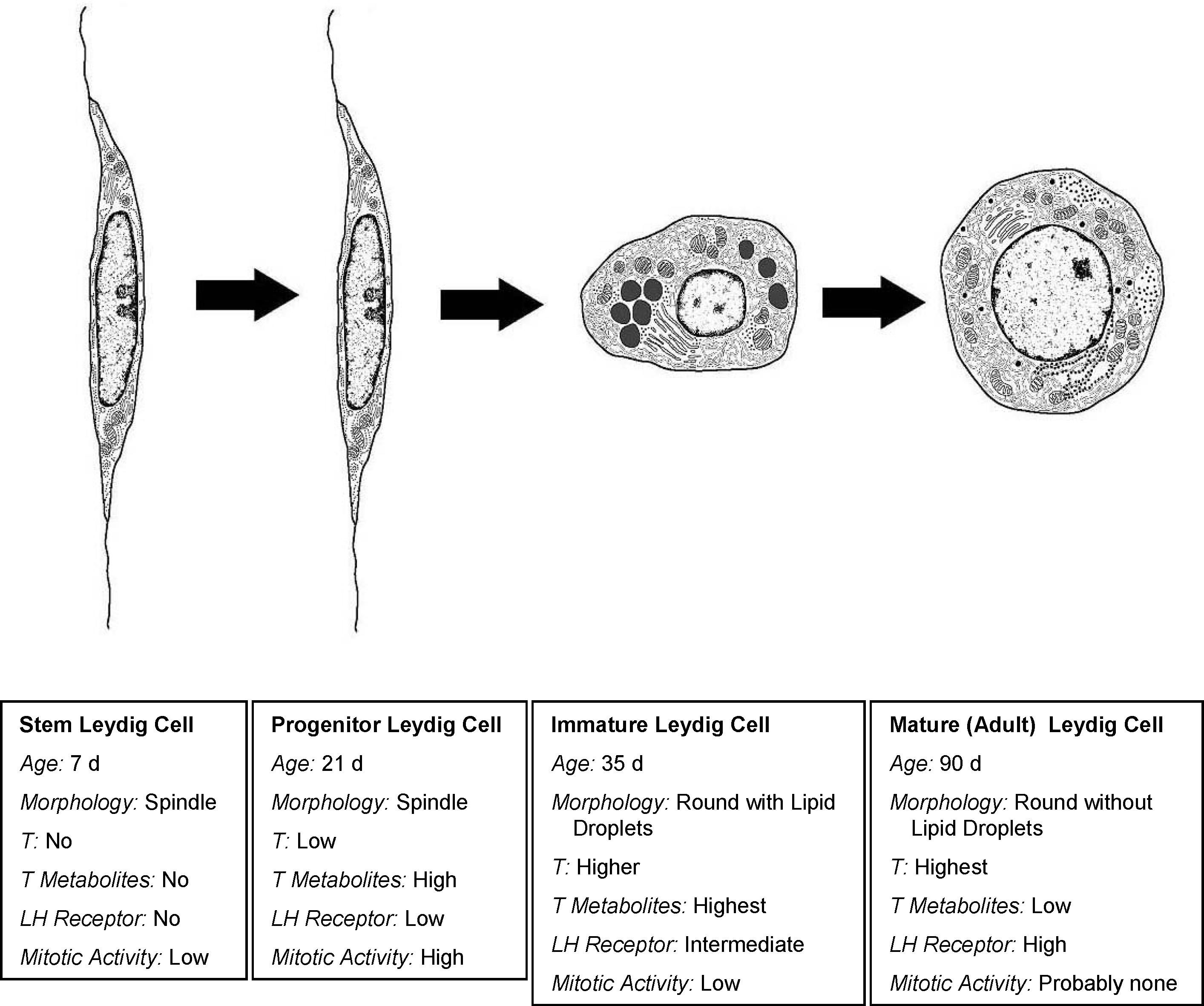
Where Do Adult Leydig Cells Come From?1

Ibuprofen and Leydig cell steroidogenic function. (A–C) Representative
Biomedicines, Free Full-Text
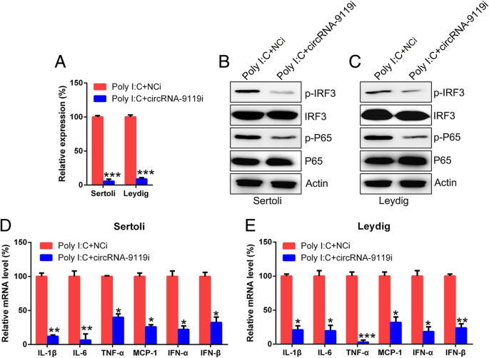
CircRNA-9119 suppresses poly I:C induced inflammation in Leydig and Sertoli cells via TLR3 and RIG-I signal pathways, Molecular Medicine
Recomendado para você
-
 Teste de Velocidade: Internet Vivo Fibra RJ 300 Megas - Vale a Pena?15 julho 2024
 -
 Vivo realiza testes para diferentes aplicações do 5G no Rio de Janeiro15 julho 2024
 -
 Medlevensohn realiza ao vivo Teste de PSA para câncer de próstata - LabNetwork15 julho 2024
 -
 Blogueiro De Beleza Ao Vivo Maquiagem Diária Escovando O Teste Na Mão Usando O Telefone Celular No Tripé. Mulher Asiática Atraente, Influenciador Da Internet, Ou Vlogger Streaming Para Criar Conteúdo On-line. Foto15 julho 2024
 -
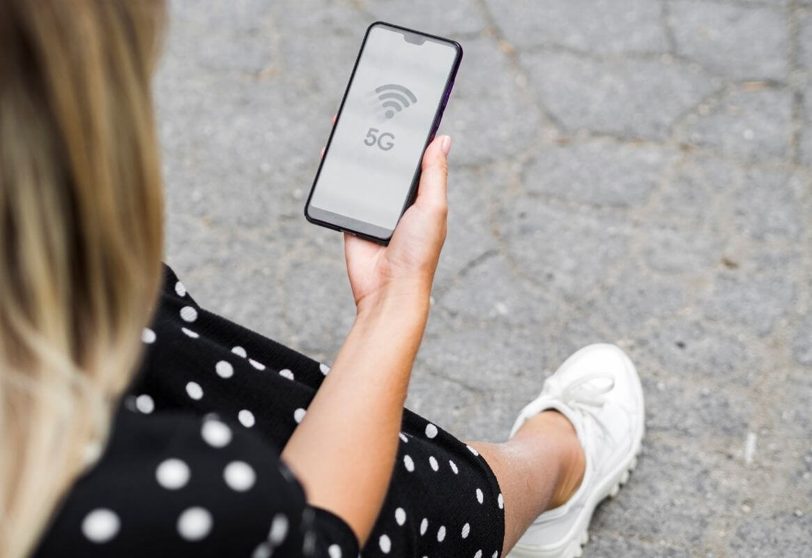 Vivo e Nokia realizam teste 5G em ondas milimétricas no Rio de Janeiro15 julho 2024
 -
/i.s3.glbimg.com/v1/AUTH_da025474c0c44edd99332dddb09cabe8/internal_photos/bs/2023/J/I/RRc9uPSAu4dWxgtdUqlg/tim.webp) TIM cria 'test-drive' para atrair clientes das rivais Claro e Vivo15 julho 2024
TIM cria 'test-drive' para atrair clientes das rivais Claro e Vivo15 julho 2024 -
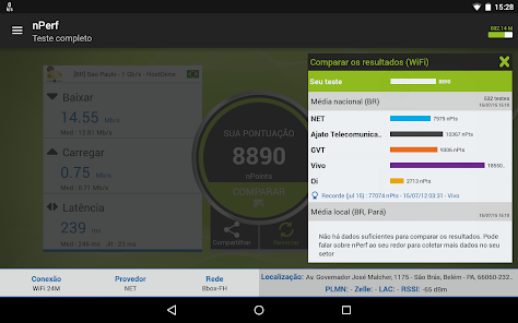
Teste velocidade 4G 5G WiFi – Apps no Google Play15 julho 2024
-
 Vai na Fé: Falsificando teste, Ben quase é pego por ligação: Dinheiro vivo15 julho 2024
 -
![TÓPICO DEDICADO] - Qual a velocidade da sua internet e a velocidade reportada no teste?, Page 416](https://www.speedtest.net/result/14336974508.png) TÓPICO DEDICADO] - Qual a velocidade da sua internet e a velocidade reportada no teste?, Page 41615 julho 2024
TÓPICO DEDICADO] - Qual a velocidade da sua internet e a velocidade reportada no teste?, Page 41615 julho 2024 -
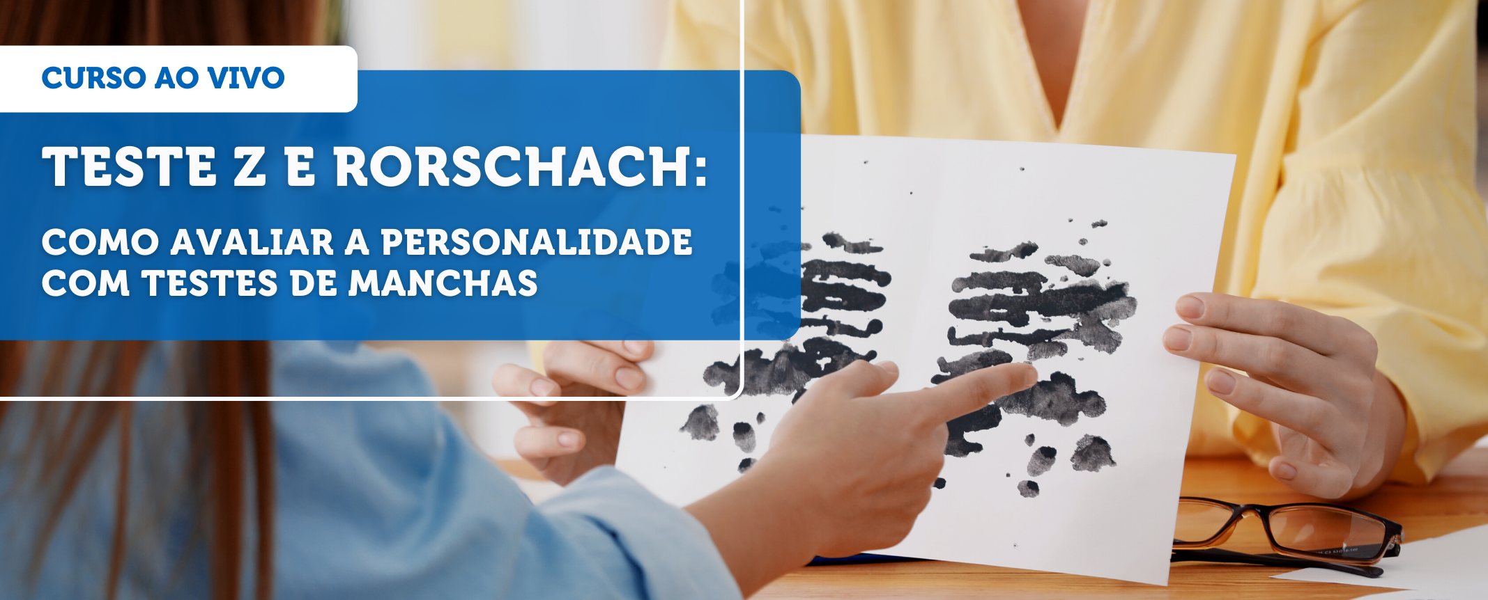 Rorschach e Teste Z: como avaliar a personalidade com testes de manchas - Grupo Educativa15 julho 2024
você pode gostar
-
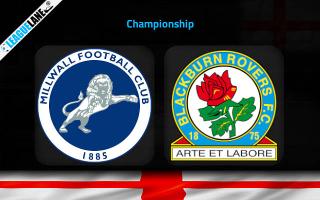 Millwall vs Blackburn Rovers Prediction, Head-To-Head, Live Stream Time, Date, Team News, lineup news, Odds, Stats, Betting Tips Trends, Where To Watch Live Score English League Championship 2023 Telecast Today Match Details –15 julho 2024
 -
/i.s3.glbimg.com/v1/AUTH_59edd422c0c84a879bd37670ae4f538a/internal_photos/bs/2019/m/n/pPTh6yQCC5TYnyIDwZ1A/corpo-recuperado.jpg) Equipes resgatam corpo em destroços de avião onde estava o jogador Emiliano Sala, Mundo15 julho 2024
Equipes resgatam corpo em destroços de avião onde estava o jogador Emiliano Sala, Mundo15 julho 2024 -
 Why your Twitch follower count means $#!%, and why Follow for Follow is a waste of time. Gameonaire15 julho 2024
 -
 Hikari no Ou – 02 – Random Curiosity15 julho 2024
 -
 Sasuke vs Udon15 julho 2024
 -
:strip_icc()/i.s3.glbimg.com/v1/AUTH_08fbf48bc0524877943fe86e43087e7a/internal_photos/bs/2023/q/F/Y27Ii9TOCDSFGNCvE9aA/350787253-238031485528151-7415373187829982385-n.jpg) Denver Nuggets x Miami Heat ao vivo: onde assistir ao jogo da NBA online15 julho 2024
Denver Nuggets x Miami Heat ao vivo: onde assistir ao jogo da NBA online15 julho 2024 -
 3D model Mario Luigi And Yoshi From Game VR / AR / low-poly15 julho 2024
 -
 O método Del Valle - Footure - Futebol e Cultura15 julho 2024
 -
 Brasileirão: como foram os últimos jogos entre Palmeiras e Santos?15 julho 2024
 -
 13 melhor ideia de Xadrez de bruxo15 julho 2024