PDF] Brain Tumor Segmentation of MRI Images Using Processed Image Driven U-Net Architecture
Por um escritor misterioso
Last updated 31 março 2025
![PDF] Brain Tumor Segmentation of MRI Images Using Processed Image Driven U-Net Architecture](https://d3i71xaburhd42.cloudfront.net/c750894747d2b3f841de55922b2b68794295de27/7-Table3-1.png)
A fully automatic methodology to handle the task of segmentation of gliomas in pre-operative MRI scans is developed using a U-Net-based deep learning model that reached high-performance accuracy on the BraTS 2018 training, validation, as well as testing dataset. Brain tumor segmentation seeks to separate healthy tissue from tumorous regions. This is an essential step in diagnosis and treatment planning to maximize the likelihood of successful treatment. Magnetic resonance imaging (MRI) provides detailed information about brain tumor anatomy, making it an important tool for effective diagnosis which is requisite to replace the existing manual detection system where patients rely on the skills and expertise of a human. In order to solve this problem, a brain tumor segmentation & detection system is proposed where experiments are tested on the collected BraTS 2018 dataset. This dataset contains four different MRI modalities for each patient as T1, T2, T1Gd, and FLAIR, and as an outcome, a segmented image and ground truth of tumor segmentation, i.e., class label, is provided. A fully automatic methodology to handle the task of segmentation of gliomas in pre-operative MRI scans is developed using a U-Net-based deep learning model. The first step is to transform input image data, which is further processed through various techniques—subset division, narrow object region, category brain slicing, watershed algorithm, and feature scaling was done. All these steps are implied before entering data into the U-Net Deep learning model. The U-Net Deep learning model is used to perform pixel label segmentation on the segment tumor region. The algorithm reached high-performance accuracy on the BraTS 2018 training, validation, as well as testing dataset. The proposed model achieved a dice coefficient of 0.9815, 0.9844, 0.9804, and 0.9954 on the testing dataset for sets HGG-1, HGG-2, HGG-3, and LGG-1, respectively.
![PDF] Brain Tumor Segmentation of MRI Images Using Processed Image Driven U-Net Architecture](https://www.mathworks.com/help/images/segment3dbraintumorsusingdeeplearningexample_01_ja_JP.png)
3-D Brain Tumor Segmentation Using Deep Learning - MATLAB & Simulink Example
![PDF] Brain Tumor Segmentation of MRI Images Using Processed Image Driven U-Net Architecture](https://www.med.upenn.edu/cbica/assets/user-content/images/BraTS/brats-tumor-subregions.jpg)
3D MRI Brain tumor segmentation, U-NET
![PDF] Brain Tumor Segmentation of MRI Images Using Processed Image Driven U-Net Architecture](https://ijisae.org/public/journals/1/article_2579_cover_en_US.png)
Patient-Specific Brain Tumor Segmentation using Hybrid Ensemble Classifier to Extract Deep Features
![PDF] Brain Tumor Segmentation of MRI Images Using Processed Image Driven U-Net Architecture](https://www.rsipvision.com/wp-content/uploads/2018/08/Joint_reconstruction_and_segmentation_2-1.png)
Deep Learning in Brain Imaging - Medical Image Analysis by RSIP Vision
![PDF] Brain Tumor Segmentation of MRI Images Using Processed Image Driven U-Net Architecture](https://www.science.org/cms/10.1126/sciadv.add3607/asset/f006810b-5ff3-4034-a144-00b49132fbcb/assets/images/large/sciadv.add3607-f1.jpg)
SynthSR: A public AI tool to turn heterogeneous clinical brain scans into high-resolution T1-weighted images for 3D morphometry
![PDF] Brain Tumor Segmentation of MRI Images Using Processed Image Driven U-Net Architecture](https://www.researchgate.net/publication/349902492/figure/fig1/AS:1024432529215497@1621255156793/Proposed-tumor-segmentation-and-classification-architecture.png)
Proposed tumor segmentation and classification architecture
![PDF] Brain Tumor Segmentation of MRI Images Using Processed Image Driven U-Net Architecture](https://onlinelibrary.wiley.com/cms/asset/85e7cbd1-a542-4c37-9d7f-16c756eeef98/ima22571-fig-0008-m.jpg)
International Journal of Imaging Systems and Technology, IMA
![PDF] Brain Tumor Segmentation of MRI Images Using Processed Image Driven U-Net Architecture](https://www.medrxiv.org/content/medrxiv/early/2022/11/04/2022.11.03.22281923/F1.large.jpg)
Comparing 3D, 2.5D, and 2D Approaches to Brain Image Segmentation
![PDF] Brain Tumor Segmentation of MRI Images Using Processed Image Driven U-Net Architecture](https://media.springernature.com/m685/springer-static/image/art%3A10.1038%2Fs41598-023-47107-7/MediaObjects/41598_2023_47107_Fig1_HTML.png)
Utilizing deep learning via the 3D U-net neural network for the delineation of brain stroke lesions in MRI image
![PDF] Brain Tumor Segmentation of MRI Images Using Processed Image Driven U-Net Architecture](https://images.prismic.io/encord/57bd343a-7e54-4653-a716-f8fbd88d1afc_image+%284%29.png?auto=compress%2Cformat&fit=max)
Guide to Image Segmentation in Computer Vision: Best Practices
brain-tumor-segmentation · GitHub Topics · GitHub
![PDF] Brain Tumor Segmentation of MRI Images Using Processed Image Driven U-Net Architecture](https://doc.pmod.com/pai/clip0247.png)
Case Studies - Application of PAI > Brain Tumor Segmentation - MICCAI Challenge
![PDF] Brain Tumor Segmentation of MRI Images Using Processed Image Driven U-Net Architecture](https://production-media.paperswithcode.com/tasks/Webp.net-resizeimage_4_VDcks9s.png)
Medical Image Segmentation
![PDF] Brain Tumor Segmentation of MRI Images Using Processed Image Driven U-Net Architecture](https://ars.els-cdn.com/content/image/1-s2.0-S2666307422000213-gr1.jpg)
Segmentation and classification of brain tumor using 3D-UNet deep neural networks - ScienceDirect
Recomendado para você
-
 How to Beat Brain Test Level 191 Walkthrough31 março 2025
 -
 Brain Test: Tricky Puzzles - Seviye 191 • Game Solver31 março 2025
 -
 brain test nível 19131 março 2025
 -
 Optical Illusion Brain Test: If you have Sharp Eyes Find the number 161 among 191 in 6 Seconds? - News31 março 2025
 -
 Solved 191 EXERCISE 27, BRAIN AND CRANIAL NERVES QUIZ 1.31 março 2025
 -
 Maker de Aplicativos31 março 2025
 -
 Brain Test 3 Level 191, 192, 193, Gameplay31 março 2025
 -
 Astronomy UV beads Solar Beads Lab Help Stop Skin Cancer Middle School Science - Classful31 março 2025
 -
 Need Movement Activities To Keep Kids Active At School? - Top Notch Teaching31 março 2025
 -
 Brain Test 4: Tricky Friends Level 191-200 Walkthrough Solution (NEW UPDATE)31 março 2025
você pode gostar
-
 Game Saints Row - Day One Edition - Xbox Series X em Promoção na31 março 2025
 -
 I Made The WORLD'S FASTEST DUCK!31 março 2025
 -
 The welcome messages for each of the four Hogwarts houses on Pottermore : r/harrypotter31 março 2025
 -
 igrodel31 março 2025
 -
 Cenário na Unreal Engine, Aluno SAGA: Samuel Cazetta31 março 2025
 -
 Sem folga: confira o calendário do futebol em 2021 e datas de estreia31 março 2025
 -
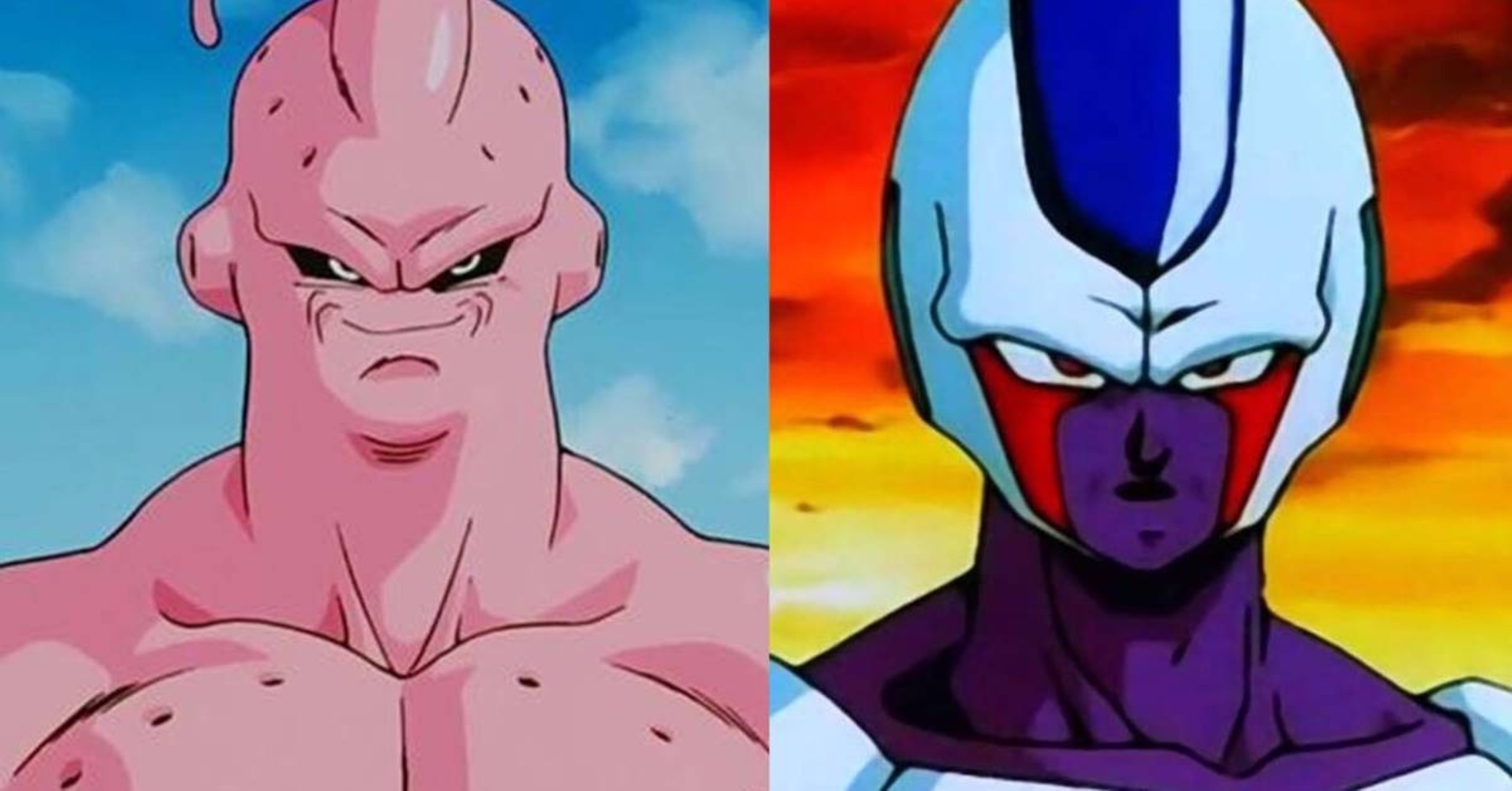 Este seria o visual de Majin Boo caso ele tivesse absorvido Cooler em Dragon Ball Z - Critical Hits31 março 2025
 -
 HOW TO GET DENJI, POWER, AKI, MAKIMA AND HIMENO IN ANIME ADVENTURES!31 março 2025
 -
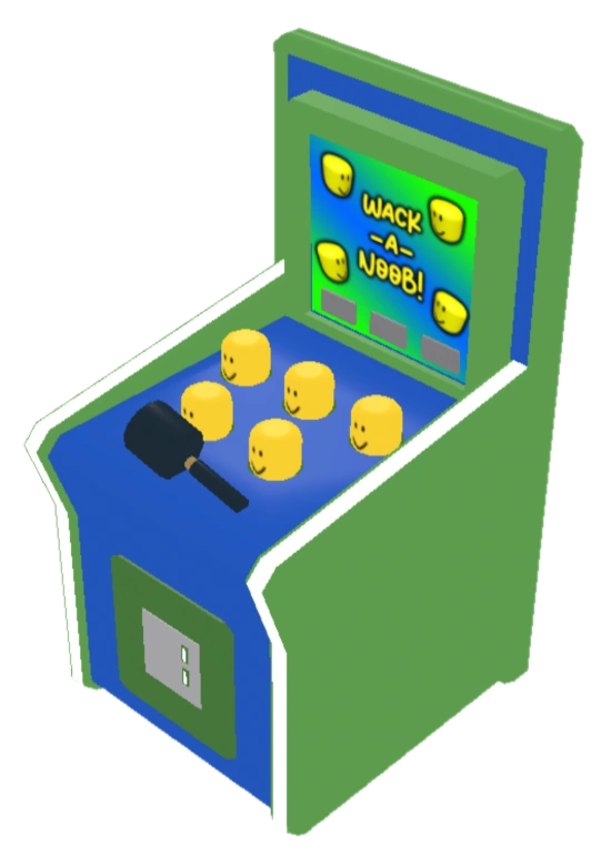 Wack A Noob, Roblox Arcade Empire Wiki31 março 2025
 -
 The Witcher': lo que sabemos de la temporada 4, que cambia a Henry31 março 2025